This isn’t my first post about the relationship between the psoas and the diaphragm.
The essential connection between the psoas and the diaphragm has a profound influence on how well our bodies function.
Considering the diaphragm as the main muscle of breathing and the psoas as the main muscle of walking, you can’t take a step that doesn’t involve the diaphragm or a breath that doesn’t involve the psoas.
And if you don’t have good posture, neither the diaphragm nor the psoas will work correctly.
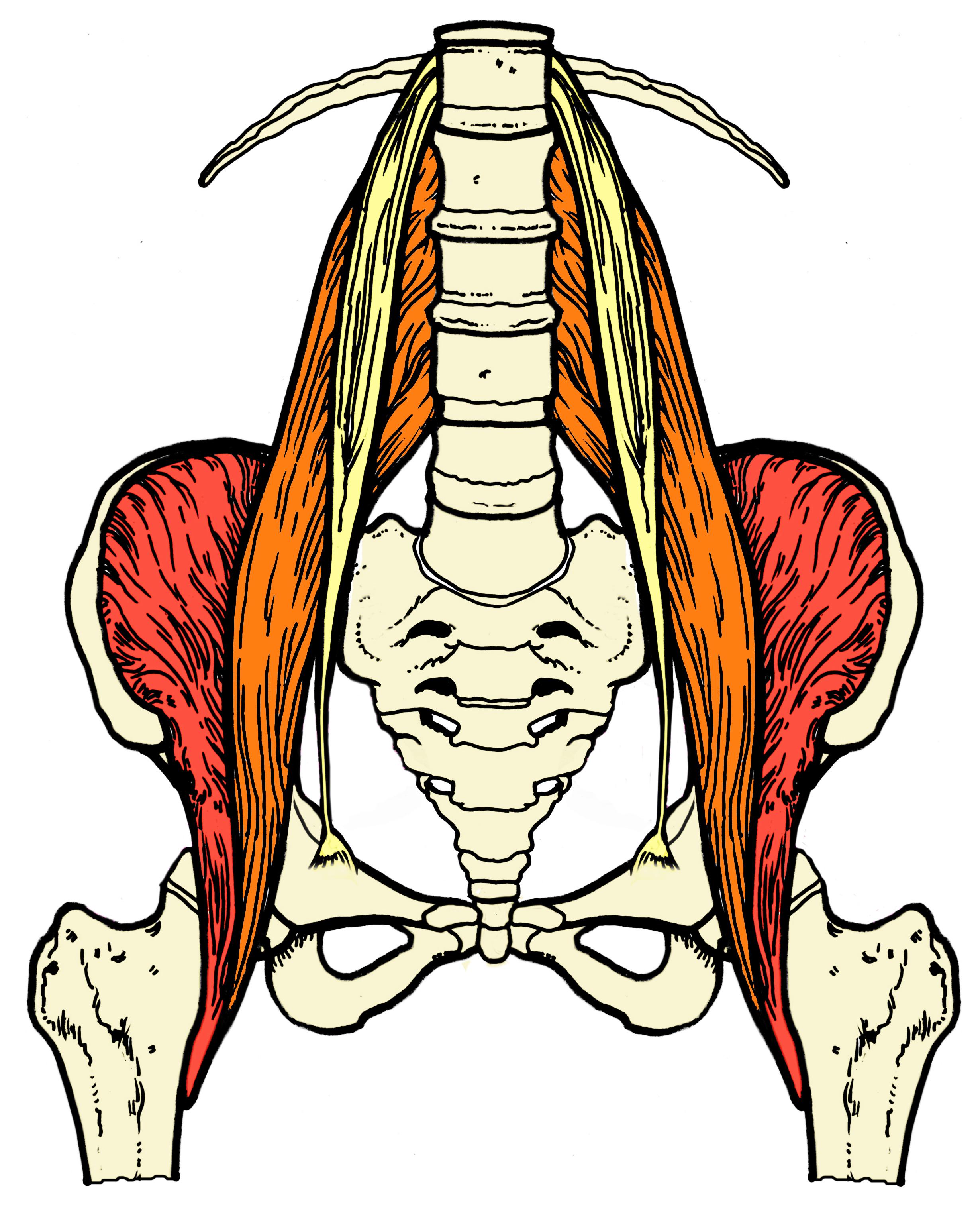
The iliopsoas muscle group is composed of the iliacus, psoas major and psoas minor.
The iliacus and psoas major share a common tendon that attaches on the back half of the inner thigh on a knob of bone called the lesser trochanter. Sharing a common tendon means that they share certain functions.
While both the psoas major and the iliacus act as flexors of the hip the psoas major has other responsibilities as well.
It is involved in external rotation of the thigh, it stabilizes the connection of the leg into the pelvis where the femoral head inserts into the acetabulum, and also assists to stabilize and erect the lower spine.
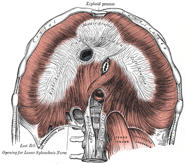
The diaphragm is the main muscle of respiration and it is different than most muscles in the body.
For one, it has openings (hiatuses) that allow for structures to pass through from the upper to lower trunk. The esophagus, abdominal aorta, vena cava all traverse the diaphragm to get from the chest to the abdomen. You don’t find a lot of muscles with stuff passing through them.
As a dome like shape at the base of the ribcage its origin is at the back along the vertebrae of the spine and at the front on the ribs and the sternum.
There is a central tendon that acts as an insertion point for the diaphragm muscle.
When we inhale and air is pulled into the lungs, the contraction of the diaphragm pulls this central tendon down allowing for air to inflate the lungs.
While the origins and insertions that I just listed suggest two separate muscles here are a few ways the diaphragm and psoas are intricately linked.
The median arcuate ligament is a ligament under the diaphragm formed by the right and left crura of the diaphragm and passes directly over the psoas major.
The diaphragm has two crura, or tendons, that attach the diaphragm to the vertebral column.
Just as the psoas major attaches along the lumbar spine, the crura of the diaphragm attach there as well. The right crus attach on the upper three lumbar vertebrae and the left crus attaches on the top two.
No muscles work in isolation and everything in the body is connected—in some ways more literally than others.
A good working relationship of the psoas and the diaphragm allows for them to function as designed and is integral to having a body that functions well and ages gracefully.
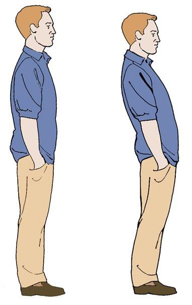
Above you’ll find my two standard pictures of posture.
From my perspective the good posture on the left is embodied by few, and the poor posture on the right is the default alignment for the majority of the planet.
For what it is worth, I think most people lean back like the picture on the right though most people do not perceive themselves that way (which is the subject of a few dozen blog posts).
The good posture picture on the left gives the psoas and the diaphragm a fighting chance to work together.
Two alignment features required for the harmonic convergence of the diaphragm and psoas are:
- The legs must be directly under the pelvis so the psoas can successfully stabilize the connection of the leg to the pelvis.
- The base of the pelvis, ribcage and floor need to be parallel to each other.
Setting the leg correctly into the hip socket and leveling the base of the rib cage allows these tacitly connected muscles to work as a unit or team driving the body to heights of functional efficiency.